
We offer BULK-BILLING
Renal Artery Duplex Ultrasound
Fasting
For this scan it is best not to eat or drink (except water) for at least four hours before your ultrasound to reduce interference from bowel gas.
What is fibromuscular dysplasia?
The renal arteries are the blood vessels which supply blood to your kidneys. It is important that the kidneys get a good supply of blood so they can function well. Unfortunately, sometimes people develop narrowing in these arteries and this is called atherosclerosis. The atherosclerosis is made up of fatty material which can become calcified and hard. In more rare cases some people can develop a different type of narrowing where the inner layer of the artery wall becomes inflamed, this is called fibromuscular dysplasia.
Why do we scan renal arteries?
If you develop atherosclerosis in your renal arteries it is possible this can result in a lack of blood to your kidneys and this is known as ischemia. Often you will be asked to get a scan of your renal arteries if you have persistent high blood pressure that won’t improve despite taking medication. This is because the kidneys play a role in blood pressure regulation. It is important to identify renal artery stenosis in patients with persistent high blood pressure as the cause can often be treated.
How is the ultrasound performed?
The vascular sonographer will use the ultrasound machine to look at the renal arteries in your abdomen. For this scan it is best not to eat or drink (except water) for at least four hours before your ultrasound to reduce interference from bowel gas. If you require food for medication then that is fine to eat. During the scan you will lie down on the bed. The sonographer will use a transducer and gel to get an image of the arteries in your abdomen. Sometimes they will have to press firmly in order to get good views of the artery. During the scan you will need to turn on to your sides for optimal views of the kidneys.
The scan usually takes about 20-30 minutes, after this the sonographer will complete their diagram and report for the referring doctor.
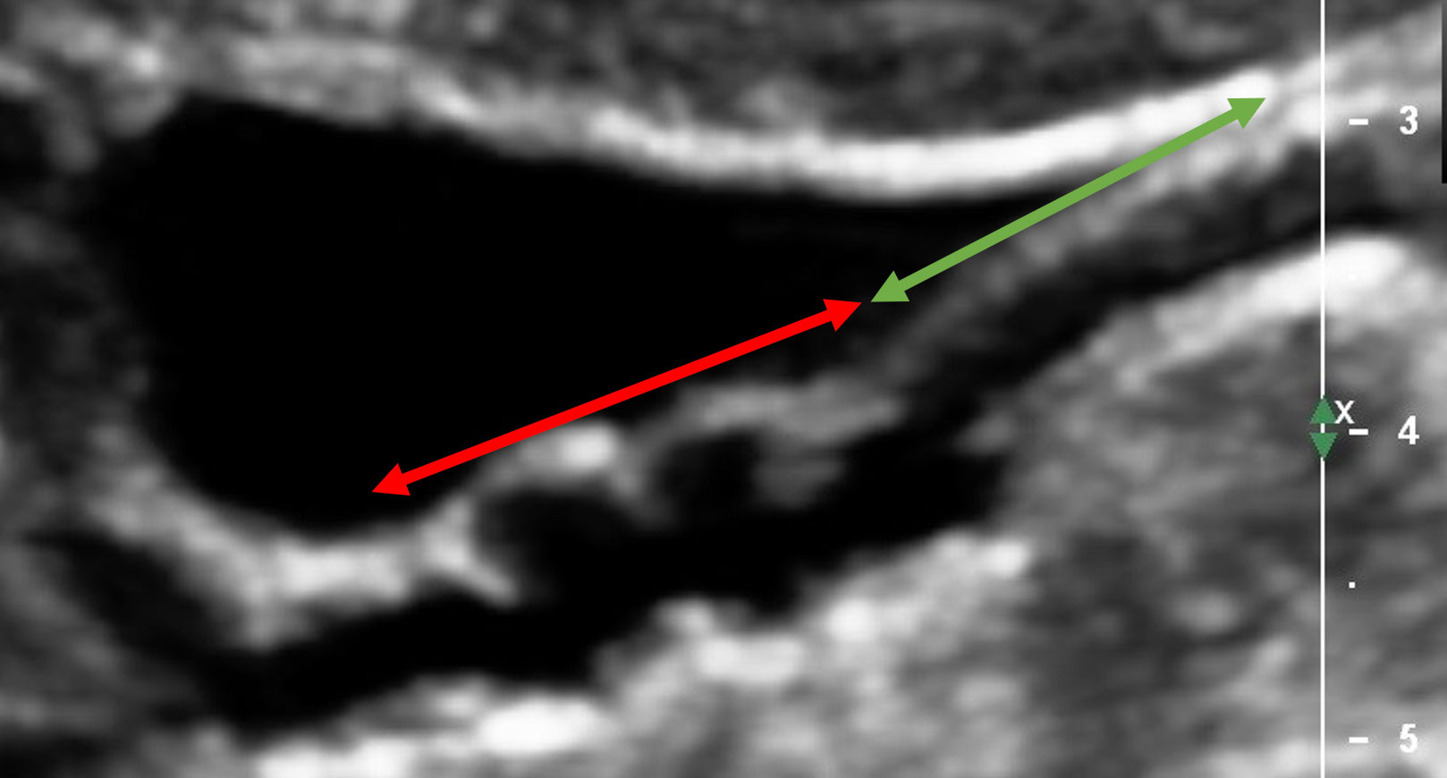
Figure 1
Evidence of FMD in the distal end of a right renal artery. The segment of proximal renal artery appears normal with no wall changes (area below the green arrow). The distal segment demonstrates the "beading" with significant wall changes (area below the red arrow)
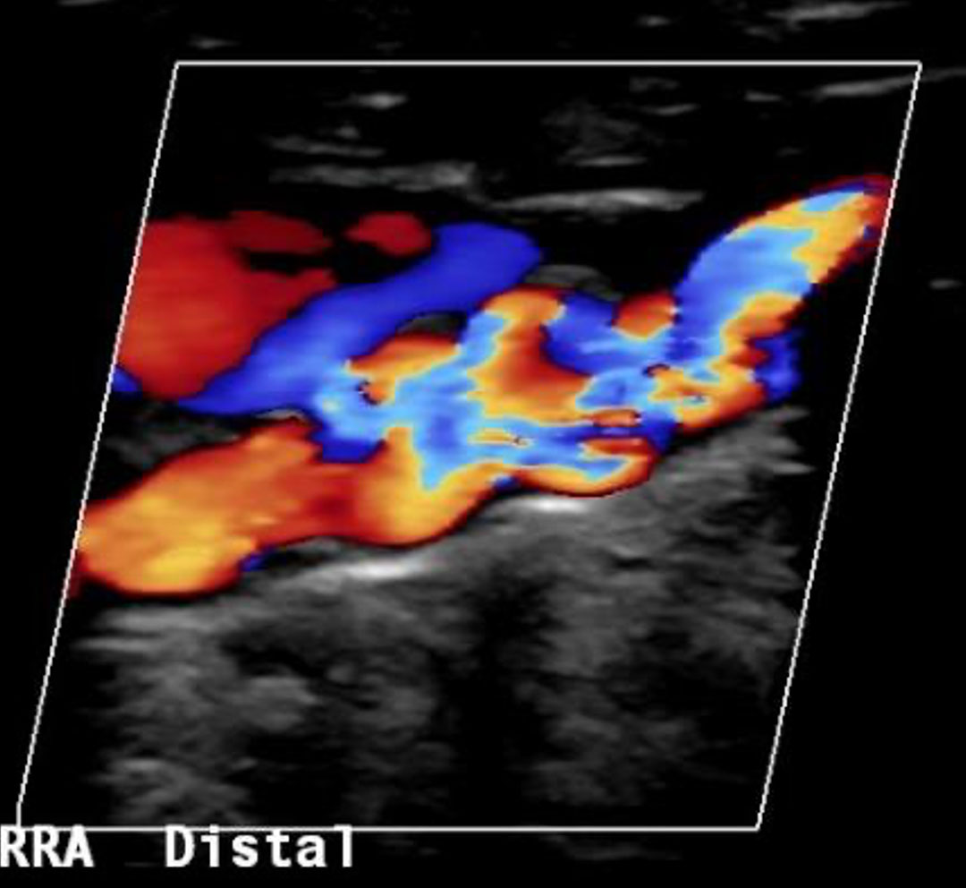
Figure 2
This shows the same segment of artery as in figure 1, the colour demonstrates the flow of blood, we can see the turbulent movement of blood through the "beading" segment of artery.
For appointments and enquiries:
Monday - Friday: 8:00am to 5:00pm
Fax: (02) 9182 7533