
We offer BULK-BILLING
Peripheral Artery Duplex Ultrasound
What is peripheral arterial disease?
The peripheral arteries are the blood vessels that transport oxygen in the blood from your heart to the muscles in your limbs. Narrowing in the arteries of the arms or legs is known as peripheral artery disease. It is more typically a problem in the legs although it can occur in the arms as well.
This narrowing is made up of fatty material which can become calcified and hard and is called atherosclerosis. If you develop atherosclerosis in the arteries of your legs, and it becomes severe enough, this can result in a lack of blood to the muscles in your legs. This may cause pain, discomfort or tiredness in your muscles of your lower legs or feet when you walk. This is known as intermittent claudication, may resolve quickly when you rest but resume when you start walking again.
As arterial disease progresses symptoms can also worsen. In some cases, in addition to pain with walking, you may also get pain in your legs or feet at night which causes you to wake up. In more extreme cases you can have what is called “critical limb ischaemia”. Symptoms often include severe pain in the legs and feet, non-healing wounds or gangrene.
How is the ultrasound performed?
Using the ultrasound machine, the vascular sonographer will use a transducer and gel and then move that along your arms or legs. They can determine how severe the atherosclerosis is by using its visual appearance and the speed that flow has to travel to get through the narrowed areas. They can also see if the artery has become completely blocked. You may have previously had stents inserted or bypass grafts, the sonographer can see these and can see if there is any new narrowing or blockage.
Once they have followed the all the major arteries in your arms or legs, the sonographer will create a provisional report for the referring doctor. This vascular ultrasound scan and your clinical history will help him to determine an appropriate treatment plan to improve your symptoms.
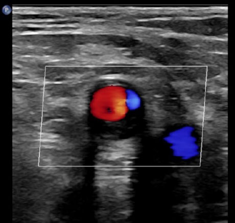
Figure 1
A common femoral artery with less than 50% stenosis
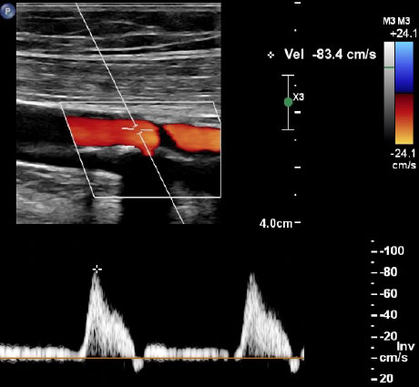
Figure 2
A superficial femoral artery with a mild calcified area of atherosclerosis
For appointments and enquiries:
Monday - Friday: 8:00am to 5:00pm
Fax: (02) 9182 7533