
We offer BULK-BILLING
Arteriovenous Fistula Surveillance
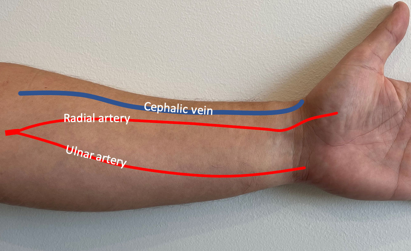
What is the arteriovenous fistula?
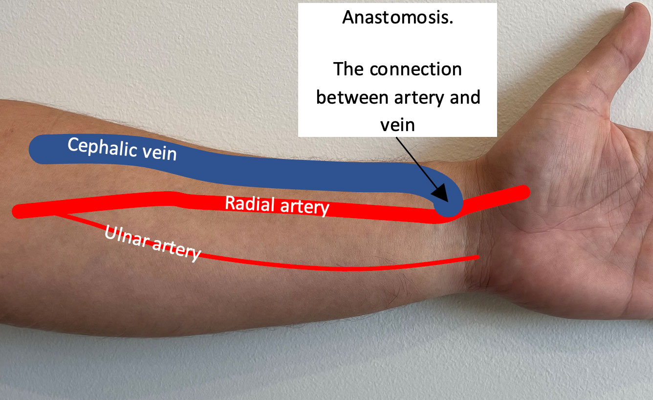
Chronic kidney disease patients who have to undergo haemodialysis will usually do so using their fistula. The arteriovenous fistula is a surgically created connection between an artery and a vein. Typically it is formed in the arm, either at the wrist or near the elbow, however some people do require their fistula to be formed in the leg in certain circumstances.
The arteriovenous fistula is essentially a shortcut for the blood flow and as a result it develops into a high flow pathway of blood. Usually the artery in the forearm (called the radial artery) will have a blood flow of less than 100ml/min. Once the surgeon has connected the vein to the artery we expect the fistula to “develop”. This means that the size of the vein will increase significantly, usually to a minimum of 5mm, and the amount of blood flow should also increase significantly, ideally to at least 500-1000ml/min.
Why does the fistula need ultrasound surveillance?
Because the fistula is formed using a small calibre artery and small calibre vein, the connection between the two vessels is also very small. Because it is so small it is possible that it can restrict the flow of blood through the fistula, this may stop the fistula from developing. If it fails to develop then it cannot be used for haemodialysis.
It is important that the fistula is routinely checked to ensure that it has not developed any narrowing or other issues that could affect the function of the fistula. If there is any significant flow limiting problem then your kidney doctors and the vascular surgeon need to know as soon as possible. Routine surveillance of the fistula using ultrasound is a safe and quick way to ensure that the fistula function is maintained.
Use navigation slider to see before and after arteriovenous fistula formation surgery.
LEFT - Before fistula formation RIGHT - After fistula formation
What happens during the scan of the fistula?
Sometimes you will need to remove your shirt so that the sonographer can check the veins in the entire arm up to your neck. Or if your fistula is in your leg then you will need to remove your trousers/pants. You do not need to remove underwear.
The sonographer will put gel on your arm (or leg) and run the transducer along your arm. They will map the fistula and note any narrowed or enlarged areas (typically at the needling sites) as well as measure the blood flow in the artery and in the vein. This scan will usually take around 20 minutes.
For appointments and enquiries:
Monday - Friday: 8:00am to 5:00pm
Fax: (02) 9182 7533