
We offer BULK-BILLING
Fibromuscular Dysplasia Duplex Ultrasound
What is fibromuscular dysplasia?
Fibromuscular dysplasia is a rare disease which affects the arteries. Unlike narrowing or blockages in the artery caused by the buildup of atherosclerosis (a fatty material which can become calcified and hard), fibromuscular dysplasia is narrowing that results from the arterial wall itself becoming enlarged, thickened and irregular in shape.
What are the symptoms of fibromuscular dysplasia?
As fibromuscular dysplasia can affect arteries in different areas of the body the symptoms can be quite variable. High blood pressure is commonly a symptom in patients who have fibromuscular dysplasia in their renal arteries. Headache, tinnitus and dizziness are commonly reported where patients have fibromuscular dysplasia in their carotid arteries. Other transient ischaemic attack (mini-stroke) symptoms can also occur with fibromuscular dysplasia.
What is the sonographer looking for?
Whenever you have an ultrasound scan of you abdominal or peripheral arteries the sonographer will be looking for changes in the blood flow and any abnormalities of the artery wall. If your doctor is concerned about the possible presence of fibromuscular dysplasia they will usually ask for you to have a carotid artery duplex ultrasound or renal artery duplex ultrasound, however in some cases they may ask the sonographer to look in other areas of your body.
The sonographer will perform the normal ultrasound scan but will ensure that they specifically note any wall changes that they find. The images below demonstrate fibromuscular dysplasia wall changes that the sonographer will be looking for. These images are of a renal artery (the artery taking blood to the kidney). The wall changes are often described as looking like a “string of beads”.
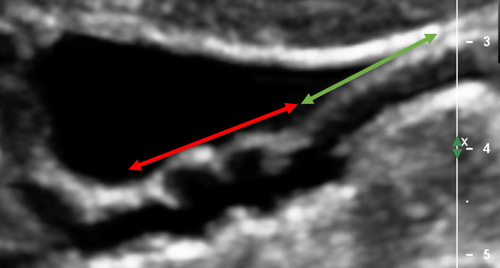
Figure 1
Evidence of FMD in the distal end of a right renal artery. The segment of proximal renal artery appears normal with no wall changes (area below the green arrow). The distal segment demonstrates the "beading" with significant wall changes (area below the red arrow)
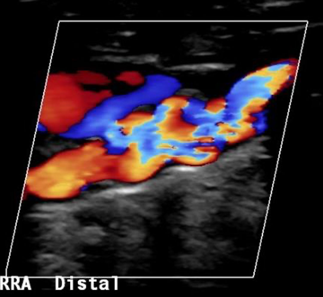
Figure 2
This shows the same segment of artery as in figure 1, the colour demonstrates the flow of blood, we can see the turbulent movement of blood through the "beading" segment of artery.
For appointments and enquiries:
Monday - Friday: 8:00am to 5:00pm
Fax: (02) 9182 7533