
We offer BULK-BILLING
Carotid and Vertebral Artery Ultrasound
What are the carotid arteries?
The carotid arteries are the blood vessels which take oxygenated blood from your heart to your brain.
Unfortunately, sometimes people develop narrowing in these arteries and this is called atherosclerosis. The atherosclerosis forms plaques which are made up of fatty material which can become calcified and hard.
If you develop atherosclerosis in your carotid arteries it is possible this can result in reduced blood flow to your brain. Sometimes plaque can break off and travel up towards the smaller arteries in the brain. If this blocks the artery, the surrounding tissue will be starved of oxygen, this is known as ischemia.
What is a TIA?
Transient ischemic attack (TIA) or stroke are often the terms used to describe the type of ischemia that has occurred. A TIA is a temporary lack of blood reaching the brain and lasts less than 24 hours. Often symptoms of a TIA or stroke will include weakness or numbness of one side of your face or body, losing the ability to speak or visual changes in one eye.
How is the ultrasound performed?
Using ultrasound your vascular sonographer is able to look inside the artery to see how much atherosclerosis has developed in your carotid arteries. The narrowing that is caused by atherosclerosis results in a high-speed flow of blood through the carotid arteries, a bit like squeezing the end of a hose pipe, and it is this fast flow that can disturb the atherosclerosis which has developed. Using the speed of the blood flow along with its visual appearance the sonographer is able to grade the narrowing that has developed and this will help Dr Theivendran plan your treatment.
During the scan you will lie down on the bed. The vascular sonographer will use a transducer and gel to take images of the arteries in each side of your neck. You will need to turn your head to the left or right depending on which side they are looking at. The scan usually takes about 10-20 minutes, after this the sonographer will complete their diagram and report for the referring doctor.
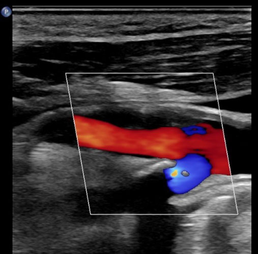
Figure 1
An internal carotid artery in longitudinal view, less than 50% stenosis
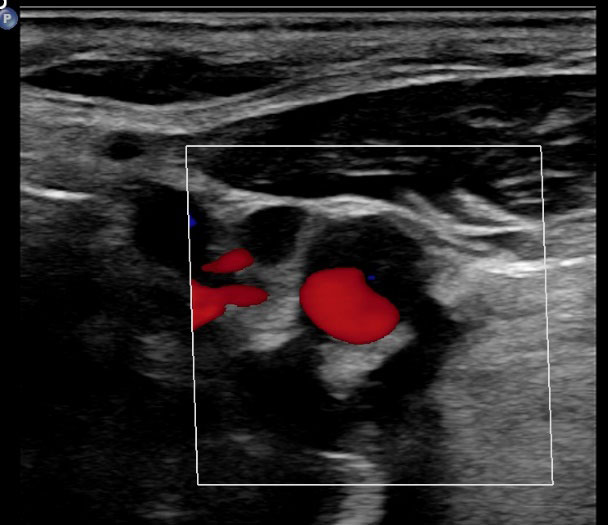
Figure 2
An internal carotid artery in cross-section, less than 50% stenosis
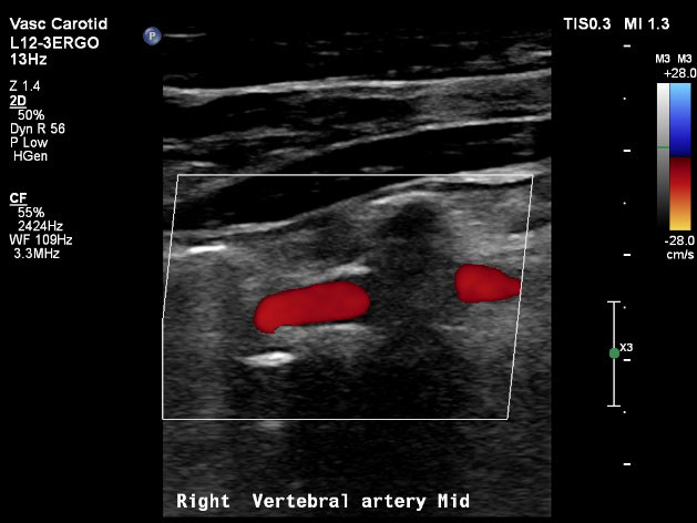
Figure 3
A vertebral artery with antegrade colour flow (toward the brain)
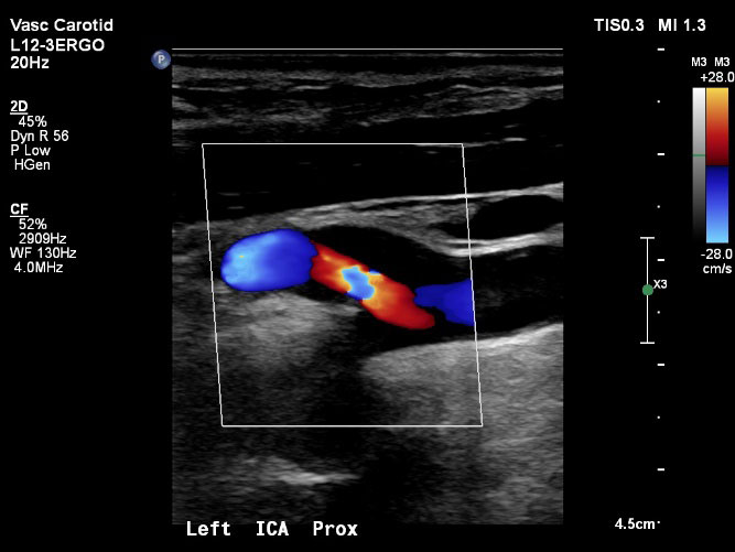
Figure 4
An internal carotid artery with a moderate to severe stenosis (70-79%)
For appointments and enquiries:
Monday - Friday: 8:00am to 5:00pm
Fax: (02) 9182 7533